
Gastroschisis is an abdominal wall defect in the fetus, affecting as many as 1 in 2000 pregnancies and growing in prevalence worldwide (Lepigeon et al, 2014). It occurs during the first trimester, and is caused when loops of bowel herniate outside the abdominal cavity in the amniotic fluid. This places the fetus at an increased risk of further complications, such as preterm labour, intra-uterine growth restriction (IUGR), fetal death in utero, bowel atresia, ischemia and volvulus (Carnaghan et al, 2016). The bowel continues to grow and develop outside the abdomen (Moore-Olufemi et al, 2015) until the fetus is born. At this point, immediate specialist intervention is needed because the exposed bowel places the neonate at risk of dehydration, hypothermia and infection (Lepigeon et al, 2014).
The contemporary literature acknowledges an increase in the global prevalence of gastroschisis in the past 30 years; however, a recent study suggests that there has been a significant stabilisation of figures, and in fact, a slight decrease has been noted (Allman et al, 2016). Research conducted in the USA found that the prevalence rate affected all demographics of age and ethnicity; however, Indigenous women in North America, Hawaii and the Pacific had higher rates of babies born with gastroschisis (Rocha et al, 2014).
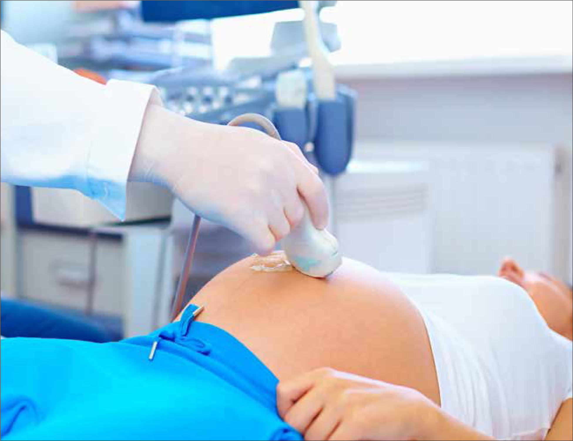
Major advances in diagnostic ultrasonography in the past 15 years have resulted in more antenatal ultrasounds showing earlier diagnosis of fetuses with gastroschisis (Faugstad et al, 2014). Ultrasonography holds a powerful role in the diagnosis, monitoring and identification of potential predictors in gastroschisis. This includes the use of the amniotic fluid index (AFI), which can be predictive of the neonate's later total parenteral nutrition requirements, or evidence of stomach herniation, which can be predictive of intra-uterine fetal death in the late third trimester (Overcash et al, 2014; Moore-Olufemi et al, 2015).
Gastroschisis can commonly be detected through routine ultrasound in the first and second trimesters, and upon serial ultrasounds following diagnosis. Any findings associated with fetal distress, death, or poor neonatal outcomes such as dilated bowel loops, IUGR, small for gestational age, or stomach and bladder herniation, provide clinicians with the opportunity for early intervention and birth (Goetzinger et al, 2014; Brown et al, 2015).
Despite the advance of ultrasonography, and in line with contemporary prevalence rates, the overall mortality rate for gastroschisis remains between 5–10% (Overcash et al, 2014; Perry et al, 2017). This figure is strongly influenced by the extent of bowel pathology in utero and the immediacy of paediatric and surgical intervention at birth (Baud et al, 2013). Fetal death can occur in the late third trimester for unknown reasons; therefore much of the research suggests birth at 37 weeks' gestation to mitigate this risk. Lakhoo (2012) argues that birth at 37 weeks can also reduce the risk of sepsis, bowel damage and prolonged neonatal hospital stay. There is much debate ongoing as to benefits of this advice, and also as to whether induction, spontaneous labour, caesarean section or vaginal birth is best practice.
‘The pregnant woman should not be viewed as separate to this complex condition, as the significant impact of serial monitoring, induced or emergency birth, and an ongoing psychosocial burden that extends into the child's early life can affect both the woman and her family’
Analysis of the literature
Early induction versus spontaneous labour has shown no bearing on neonatal outcomes, and birth at 37 weeks has been associated with adverse outcomes (Overcash et al, 2014). Elective caesarean section at 38 weeks appears to be the most cost-effective method, with the fewest interventions performed on the neonate (Harper et al, 2015), although despite preventing adverse outcomes, the majority of the available literature suggests that it provides no advantage over vaginal birth (Tarca et al, 2015). Nevertheless, as these debates continue, it needs to be recognised that 50% of all gastroschisis cases will need an emergency caesarean section, due to the high incidence of fetal distress in utero.
The contemporary research can be found to agree unanimously on the multidisciplinary approach needed in the management of gastroschisis; involving obstetricians, neonatologists, paediatricians and paediatric surgeons (Lakhoo, 2012). This also suggests that parents are faced with more information, choice and decisions than ever before, which can create problems as well as solving them.
However, the literature is yet to describe responses to potential ethical dilemmas where parents could refuse serial ultrasounds, or opt for a particular birthing practice beyond the theoretical boundary of 37 weeks' gestation.
There is also a significant lack of midwifery-led research in the literature, particularly on the distress that the pregnant woman and her partner undergo antenatally through to the postnatal period. The literature that is available is mostly medical-led, objective and quantitative, focusing on diagnostics and interventions that fit within typical medical models and outlooks.
One small study set out to address parental concerns around gastroschisis, providing clinical statistics and guidelines with complex medical jargon as a response to these concerns (Lepigeon et al, 2014). There is a wide gap in the available literature where midwifery-led gastroschisis care needs to be described and informed. Ideally, midwifery-led research would serve to round and strengthen the pool of literature, which lacks a wider spectrum of thinking and approach.
Pathophysiology
The aetiology of gastroschisis is unclear and yet to be defined. It is not associated with chromosomal anomalies, and there are only associated risk factors in the development of this birth defect (Bargy and Beaudoin, 2014). The process of the defect itself also remains somewhat unclear, although through intensive ultrasound observation and embryonic studies, greater understanding has been achieved in recent years.
Gastroschisis can be seen on an ultrasound scan at approximately 8–10 weeks of gestation, usually through routine antenatal first trimester screening. It is formed when the bowel herniates into the right side of the umbilical cord. Upon an amniotic rupture in the pars flaccida of the cord, the bowel continues to herniate through the abdomen wall, outside the fetal body (Bargy and Beaudoin, 2014).
The fetal abdomen can resolve a potential herniation, with the umbilical cord stretching and filling with Wharton's jelly, and therefore an official diagnosis is not made until after 12 weeks' gestation. Gastroschisis is thought to be the failure of the abdominal wall to correct this herniation. Alternatively, if the amniotic epithelium along the umbilical cord in the pars flaccida does not rupture, this lesser herniation is considered an omphalocele and a different pathology (Bargy and Beaudoin, 2014). Bowel herniation mostly occurs to the right of the umbilical cord where the right umbilical vein is thought to be affected, which weakens or necrotises the forming abdomen wall. A rare left-sided gastroschisis is thought to occur if there is early regression of the left umbilical vein (Mandelia, 2015).
Further bowel injuries such as atresia, volvulus and ischaemia are thought to occur from fetal vascular mesenteric impairment and/or from inflammatory markers found in the amniotic fluid, in which the herniated bowel sits. This in turn produces fetal distress, increases the need for emergency caesarean section, and results in poorer neonatal outcomes (Bargy and Beaudoin, 2014). These potential bowel injuries in utero are theoretically thought to cause the high rates of fetal death that occur around 39 weeks' gestation in cases of gastroschisis.
While there is no consensus as to the specific cause of gastroschisis, the fetus with gastroschisis is consistently male, and born to women less than 19-20 years of age (Csermely et al, 2015). Teratogens that have been implicated with a higher risk of gastroschisis occurring include alcohol consumption, the use of non-steroidal anti-inflammatory drugs (NSAIDS), opioid abuse, and smoking tobacco and cannabis (van Gelder et al, 2014; Ailes et al, 2015; Simeone et al, 2015). Each of these teratogens can have significant effect on the woman's circulation and vasculature that may contribute to the vascular mesenteric impairment mechanism in the fetus, which in turn predicts a poor outcome.
In a US study, poor diets that lack essential fruits and vegetables have also been found to hold strong association with the defect in Hispanic women and those not born in the USA (Feldkamp et al, 2014). A similar study conducted in the USA also discovered that an increased exposure of the population to pesticides drew a positive correlation to gastroschisis in the infants of mothers over the age of 20 years but not under (Kielb et al, 2014). This study inferred that these women built up enough of the pesticides over time to affect their pregnancy, which therefore raised significant concerns over environmental factors that should be considered.
The midwifery role in gastroschisis
Gastroschisis is a defect that affects the fetus and later, the surviving neonate. There is no cure but only prenatal diagnosis, serial monitoring and timely surgical intervention. The pregnant woman should not be viewed as separate to this complex condition, as the significant impact of serial monitoring, induced or emergency birth, and an ongoing psychosocial burden that extends into the child's early life can affect both the woman and her family.
A collaborative, multidisciplinary approach is necessary, involving midwives, nurses, social workers, dieticians, psychologists, obstetricians, neonatologists, paediatricians and paediatric surgeons. This collaboration will be intensive and may require the midwife to act as the fulcrum; balancing and bringing the care of the woman and baby together.
Education
The midwifery role in early antenatal education is the beginning of planned care. This includes the booking visit, as research shows that the teratogens of alcohol, tobacco, cannabis, NSAIDS and opioids have direct associations with gastroschisis, specifically 3 months after conception (van Gelder et al, 2014; Ailes et al, 2015; Simeone et al, 2015). Women under the age of 20 years are also at higher risk of conceiving a baby with gastroschisis, and compounding this risk is that adolescents in this age group are not only more likely to use the above teratogens, but often poor diet in these age groups in western societies places them in a higher risk category (Feldkamp et al, 2014). As in any pre-conception care opportunities, the midwife collaborates with dieticians in the role of a primary health educator, raising awareness and prevention opportunities to women and their partners who exhibit poor diet and/or use these teratogens.
‘A multidisciplinary approach is necessary, involving midwives, nurses, obstetricians, neonatologists, paediatricians and paediatric surgeons. This collaboration may require the midwife to act as the fulcrum; bringing the care of the woman and baby together’
If the multidisciplinary team are providing antenatal screening for women beginning in the first or early second trimester, it is expected that gastroschisis will be identified on the woman's first or second ultrasound. Importantly, diagnosis is usually delayed by medical staff until after 12 weeks' gestation, which allows for any late abdominal wall herniation to resolve (Bargy and Beaudoin, 2014).
In addition, maternal blood tests taken in the second trimester can show elevated alpha-fetoprotein levels, indirectly highlighting gastroschisis (Lepigeon et al, 2014). Midwives can provide explanations of the process between identification and diagnosis to the woman in the early stages, and they may also find themselves translating some of the more complex medical terms that arise in antenatal encounters into language that the women can readily understand.
Outcomes for women in remote settings
There is a lack of research on gastroschisis and the outcomes for women in rural or remote settings who may not have access to antenatal care, especially for Indigenous populations. In remote and rural areas of most countries there is lack of access to tertiary antenatal care, with delayed presentations, higher morbidity and mortality rates—and, therefore, gastroschisis in this situation—likely to be undiagnosed until birth (Bar-Zeev et al, 2014). Higher neonatal death with gastroschisis in rural and remote areas has been reported, but this was also related to the prolonged time between birth and surgical intervention, which lent advantage to infection and sepsis rates in the neonate (Tarca et al, 2015).
If gastroschisis is identified in remote and rural areas, multidisciplinary care is usually carefully and culturally planned in order to achieve the recommended serial ultrasounds or monitoring of the woman and fetus. A clear plan of birth is also needed, in close proximity to a tertiary hospital, where surgical intervention will not be delayed. Further research is yet needed on how to achieve effective antenatal care and diagnosis in pregnancy.
Diagnosis and monitoring of gastroschisis
Upon diagnosis, serial ultrasounds are required throughout the pregnancy to monitor fetal growth, amniotic fluid volume, fetal heart monitoring and, importantly, the appearance of the bowel (Lepigeon et al, 2014). Ultrasounds are essential during the late third trimester, as this is the period when bowel atresia, volvulus, and ischemia in response to amniotic fluid are more commonly identified; any of which can potentially cause fetal distress requiring urgent birth, or fetal death. IUGR and low birth weight are also important complications to monitor throughout pregnancy, as these are closely associated with gastroschisis and can predict poor outcome for the neonate (Lepigeon et al, 2014).
The midwife can help to facilitate serial ultrasounds by explaining to the woman and her family why they are necessary, and the processes required if any added complication is discovered, being sure to specify urgent birth and treatment of the neonate. The limitations of ultrasound should also be explained, as surveillance is not curative, and ultrasound does not prevent any of the complications for which it searches. Abdomen circumference leading to IUGR diagnosis can be unreliable, as the external bowel prevents accurate measurement of the abdomen and can create high rates of false positives or negatives (Tarca et al, 2015).
If there is a discussion regarding termination of the pregnancy, specific processes need to be clarified, as well as the psychosocial impact of this choice. Comprehensive discussion is required, since the stigma of a birth defect may affect the woman's understanding of neonatal outcomes. The woman and her family need to understand that more than 90% of gastroschisis babies survive and, with medical intervention, ‘good to excellent quality of life is reported by 80% to 100%’ of those grown children (Lepigeon et al, 2014: 319).
The midwife's role during labour and birth
During birth, regardless of mode, the midwife must be extra diligent in addition to their normal labour and birth duties, to ensure that obstetricians, paediatricians, and paediatric surgeons are present to assist, or are on standby. This is because the woman will require antibiotics prophylactically and continuous fetal heart monitoring to observe for fetal distress. As the neonate may require resuscitation at birth, and as the exposed bowel at birth places the neonate at risk of rapid dehydration, hypothermia, and infection, an urgent response will also be needed (Overcash et al, 2014).
The woman will also need support for the further intervention in theatre or a neonatal intensive care unit that will necessitate a sudden separation from the neonate after birth. Ideally, preparation and pre-counselling for this event will have occurred during the antenatal period, and the midwife can also act as a liaison between the woman and neonatal departments, to ensure that regular contact and bonding can occur.
There are no research studies comparing and analysing the immediate clinical response of gastroschisis in the neonate at birth, yet anecdotally, in the authors' own clinical practice, the guidelines and policies suggest wrapping a clear, sterile drawstring bag around the exposed bowel at birth, providing neonatal resuscitation if needed, and then arranging immediate transport to a special care nursery for intravenous cannulation and preparation of surgery to return the bowel into the abdominal cavity. The timing depends on the severity of the gastroschisis.
The immediate future of the neonate through surgery, and the initiation of enteral or total parenteral nutrition feeding depends largely on the potential complications of gastroschisis detected antenatally. The midwife can encourage colostrum harvesting, which is a process involving antenatal expressing and storing of a woman's colostrum (Wszolek, 2015). This will encourage the woman's milk supply for when the neonate can tolerate enteral feeds and later breastfeeding. Early expressing can also determine successful continuation of later breastfeeding (Johnson et al, 2013).
It should also be noted that donor breast milk has been used in intensive care nurseries since the 1990s, and research has found that it can prevent necrotising enterocolitis and improve overall survival rates (Bulpitt et al, 2014). While there is no research specific to gastroschisis, the neonate is at particular risk of necrotising enterocolitis, and if the woman's breast milk supply is sufficient at the time when the neonate can cope with enteral feeds and/or breastfeeding, this would be advantageous to both the woman and her baby immediately and in the long term (Sadlier, 2013).
Conclusion
Gastroschisis has increased in prevalence and therefore more cases can be expected. While much is yet to be understood about its aetiology and embryonic processes, improvements in the surveillance of gastroschisis mean that, through serial ultrasounds in antenatal care, this complex condition's pathophysiology is becoming understood, enabling both the woman and the fetus to receive targeted multidisciplinary care. It is recommended that the midwife be supportive and an advocate in partnering with the woman through education, informed choice and multidisciplinary counselling.
Future midwifery research, with a focus on pregnancy with gastroschisis, is required, and research associated with the psychosocial impact of gastroschisis, breastfeeding, and the potential ethical considerations regarding the screening and treatment choices available to women, should also be considered. In addition, midwifery practice needs further research to inform the response to cases of gastroschisis in rural and remote settings, where there is limited access to tertiary care.